
Metformin
Athanor · official
C4H11N5 · 129.16 g/mol
IUPAC name: 2-carbamimidoyl-1,1-dimethylguanidine
Also called Glucophage, biguanide, dimethylbiguanide
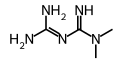
Names and identifiers
- IUPAC name
- 2-carbamimidoyl-1,1-dimethylguanidine
- Synonyms
- Glucophage, biguanide, dimethylbiguanide
+ 14 more
[14C]metformin, [14C]-metformin, SCHEMBL8944, SCHEMBL8945, GTPL4503, GTPL4779, orb1220037, orb1298860, SCHEMBL9913821, SCHEMBL10276396, SCHEMBL10325534, SY046515, NS00000333, Q19484
- Molecular formula
- C4H11N5
- Empirical formula
- C4H11N5
- Molecular weight
- 129.16 g/mol
- Exact mass
- 129.10145
- CAS number
- 657-24-9
- PubChem CID
- 152743144
- Canonical SMILES
- CN(C)C(=NC(=N)N)N
- Isomeric SMILES
- CN(C)C(=NC(=N)N)N
- InChI
- InChI=1S/C4H11N5/c1-9(2)4(7)8-3(5)6/h1-2H3,(H5,5,6,7,8)
- InChIKey
- XZWYZXLIPXDOLR-UHFFFAOYSA-N
- Molfile
Names, InChI and the identifiers from PubChem, retrieved 2026-10-14.
Calculated
- Heavy atoms
- 9
- Rings
- 0
- Aromatic rings
- 0
- Fraction sp³ carbons (Fsp3)
- 0.5
- H-bond donors
- 3
- H-bond acceptors
- 1
- Rotatable bonds
- 0 computed here · PubChem 2
- TPSA (Ų)
- 91.5
- Formal charge
- 0
- XLogP3
- -1.3 PubChem
- Complexity
- 136 PubChem
- Stereocenters
- None
- Double-bond geometry
- None specified
- Point group
- C1 of one 3D conformer with its hydrogens, by Athanor’s engine (as in the app)
Elemental analysis
- C37.20%
- H8.58%
- N54.22%
Isotope pattern
Each peak is a nominal mass with its abundance-weighted exact mass, as a percentage of the tallest, from IUPAC natural isotope abundances.
Computed from the structure with RDKit 2026.09 when this page was built, beside PubChem's own computed values from its record; where the two differ, both are shown.
Predicted
- log P
- -1.24 Crippen (Wildman and Crippen 1999)
- log S (water solubility)
- 0.14 ESOL (Delaney 2004), log mol/L
- Synthetic accessibility
- 3.21 SA score, 1 easy to 10 hard (Ertl and Schuffenhauer 2009)
Drug-likeness: Lipinski’s rule of five
- Molecular weight ≤ 500: 129.17
- log P ≤ 5 (Crippen): -1.24
- H-bond donors ≤ 5: 3
- H-bond acceptors ≤ 10: 1
Passes all four rules.
¹³C NMR shifts, predicted
Predictions, not measurements; each names its method. A measured log P, where a source reports one, is under Physical properties.
Safety
No GHS hazard classification was found in the sources.
Safety data sheets on the supplier’s page: MolCore, LGC Standards, Activate Scientific.
Always read the supplier’s safety data sheet before working with it.
Spectra
- 1H NMRHuman Metabolome Database (HMDB) (Varian · 600 MHz · Water)
- 1H-13C NMRHuman Metabolome Database (HMDB) (Bruker · 600 MHz · Water)
- GC-MSSpectraBase (DI)
- MS-MSHuman Metabolome Database (HMDB) (Positive) · Human Metabolome Database (HMDB) (Positive) · Human Metabolome Database (HMDB) (Positive) · Human Metabolome Database (HMDB) (Quattro_QQQ · Positive) · Human Metabolome Database (HMDB) (Quattro_QQQ · Positive) · Human Metabolome Database (HMDB) (Quattro_QQQ · Positive)
- LC-MSSpectraBase (Thermo Fisher Q Exactive Orbitrap · HCD)
- ATR-IRSpectraBase (Bio-Rad FTS · ATR-Film (MeCl2) (DuraSamplIR II))
Experimental spectra held by these sources; each link opens the source's own record.
Literature
Reading PubChem, OpenAlex and GSRS…
Bioactivity
Reading ChEMBL…
References
- [1]Human Metabolome Database (HMDB) · Linked only · via PubChem · retrieved 2026-10-14
- [2]SpectraBase · Linked only · via PubChem · retrieved 2026-10-14
- ·Calculated values: computed by Athanor with RDKit 2026.09 from the structure.
- ·Literature on PubChem
Data last updated 2026-10-14.